Phone: +1 (281) 968-0718
Email: sales@USComponent.com


28
Jul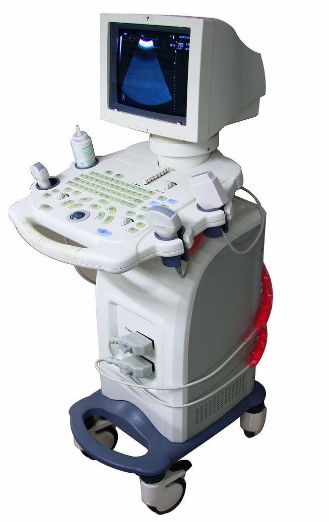
Use of IGBTs in medical ultrasonography machines

Heart specialists, neonatologists, obstetricians, urologists, gastroenterologists use ultrasound based imaging extensively for diagnosis and treatment of patients. Sound waves above the audible range of humans are called ultrasound. The choice of the ultrasound frequency for diagnostic purposes is a trade-off between image resolution and special depth. Due to the longer wavelength of the sound wave, lower ultrasound frequencies produce images with less resolution, although these penetrate deeper into the body. Normal ultrasound frequencies range from 2 to 18 MHz. A hand-held probe is used to perform Sonography that is placed and moved over the patient while viewing the image in real time. A piezoelectric transducer with a phased array is contained in the probe, which allows altering the direction and depth of the sound wave. The sound wave is reverberated from the organs inside the body at different intensities depending upon their composition and the time taken for the echo to return to the transducer specifies the distance travelled by the wave. For diagnostic purposes, this information is converted to an image.
The application of a high voltage pulse to the piezoelectric medium produces the sound wave from the ultrasound transducer. The pulse must have amplitude of over 1000 volts with a current of 20-50 amperes. Because of the short duration of the pulse, generally 0.5 microseconds, with a less operating frequency of 200-Hz, the best approach is to slowly charge a capacitor through a diode when the IGBT is off and then turn-on the IGBT for the short pulse duration to discharge the capacitor through the transducer.

USComponent is glad to offer:
- HIGH QUALITY
- FAST DELIVERY
- REASONABLE PRICE
side links
USComponent Inc.
Call us: +1 (281) 968-0718
Email: sales (at) uscomponent.com
Address: 3521 Glisten Street, Norman, OK 73072 USA

this site is ©copyright USComponent 2001-2024, all rights Reserved